IB DP Biology 2025: Unity and Diversity
A2.2 — Cell Structure
Theme A: Form and Function · Standard & Higher Level
A2.2.1
Cells as the Basic Structural Unit
Key Understanding Cells are the fundamental structural and functional unit of all living organisms.
The cell theory is one of the unifying principles of biology. Using deductive reasoning, we can predict that any newly discovered living organism will be composed of cells.
1. All living organisms are composed of one or more cells
↓
2. The cell is the basic unit of life (smallest entity that survives independently)
↓
3. All cells arise from pre-existing cells by division
A2.2.2
Microscopy Skills & Magnification
Key Understanding We can calculate magnification and real size from micrographs.
Most cells are too small to be seen with the naked eye. Microscopes magnify images, allowing biologists to study detailed ultrastructure.
Skill: Calculating Magnification
Use the MIA formula to calculate magnification, image size, or actual size.
Magnification (M) = Image Size (I) / Actual Size (A)
- I: Measure the image using a ruler (convert to µm).
- A: The real size of the specimen.
- M: How many times larger the image is compared to the specimen.
Exam Tip: Always convert your units to be the same before calculating! 1 mm = 1000 µm. 1 µm = 1000 nm.
A2.2.3
Developments in Microscopy
Key Understanding Technological advancements in microscopy have revolutionized our understanding of cell structure.
| Type | Resolution & Features | Use Cases |
|---|
| Light Microscopy |
Low resolution (~200 nm). Uses visible light. |
Viewing live cells, general tissue structure, and some organelles (nucleus). |
| Electron Microscopy (EM) |
High resolution (~0.1 nm). Uses a beam of electrons. |
Viewing dead cells at high magnification. Resolves ultrastructure (ribosomes, membranes). |
| Fluorescence Microscopy |
Uses specific fluorescent tags (fluorophores). |
Tracking specific proteins or structures within a living or fixed cell. |
| Cryo-Electron Microscopy |
Samples frozen at cryogenic temperatures. |
High-resolution 3D structures of biomolecules without the need for dyes or fixatives. |
A2.2.4
Common Cellular Structures
Key Understanding All cells, regardless of type, share certain fundamental structures required for life.
Whether prokaryotic or eukaryotic, every living cell possesses the following structural components:
Plasma Membrane
A phospholipid bilayer that acts as a selectively permeable boundary.
Cytoplasm
A water-based medium where metabolic reactions occur.
DNA
The genetic material that stores instructions for life processes.
Ribosomes
The molecular machines responsible for protein synthesis.
A2.2.5
Prokaryotic Cell Ultrastructure
Key Understanding Prokaryotes have a simple, uncompartmentalized cell structure.
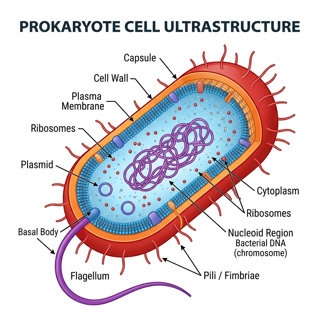
- Nucleoid: Region containing a single, continuous loop of naked DNA.
- 70S Ribosomes: Smaller ribosomes for protein synthesis.
- Cell Wall: Rigid outer layer (peptidoglycan) that maintains shape and prevents bursting.
- Plasma Membrane: Controls entry and exit of substances.
- Cytoplasm: Site of metabolic reactions.
A2.2.6
Eukaryotic Cell Ultrastructure
Key Understanding Eukaryotes possess a compartmentalized cell structure with membrane-bound organelles.
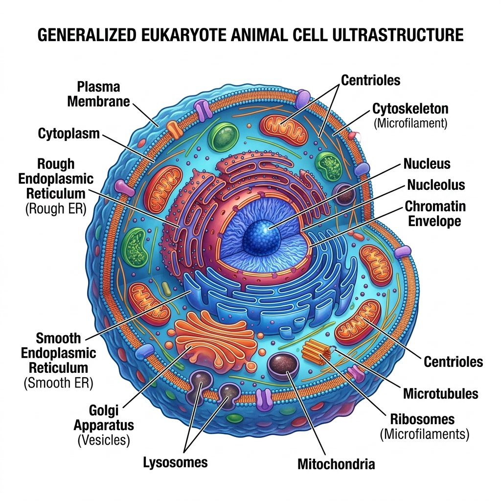
- Nucleus: Contains linear DNA associated with histones; enclosed in a double membrane with pores.
- 80S Ribosomes: Larger ribosomes bound to the Rough ER or free in cytoplasm.
- Mitochondria: Site of aerobic respiration (ATP production).
- Endoplasmic Reticulum (RER & SER): Synthesis and transport of proteins (RER) and lipids (SER).
- Golgi Apparatus: Modifies, sorts, and packages proteins for secretion.
- Vesicles & Vacuoles: For transport and storage.
- Cytoskeleton: Structural support and movement.
A2.2.7
Life Processes in Unicellular Organisms
Key Understanding A single cell must carry out all the functions of life.
Application: Paramecium and Chlamydomonas
Investigate the life processes (MR SHENG) in unicellular organisms like Paramecium (heterotroph) or Chlamydomonas (autotroph) as examples of independent living cells.
| Function (MR SHENG) | Description |
|---|
| Metabolism | Enzyme-catalyzed chemical reactions in the cytoplasm. |
| Reproduction | Producing offspring (e.g., binary fission). |
| Sensitivity | Responding to internal and external environmental stimuli. |
| Homeostasis | Maintaining a stable internal environment (e.g., osmoregulation via contractile vacuoles). |
| Excretion | Removal of metabolic waste products. |
| Nutrition | Obtaining food for energy and growth (autotrophic or heterotrophic). |
| Growth | Increasing in size or dry mass over time. |
A2.2.8 / A2.2.9
Eukaryotic Variations & Atypical Cells
Key Understanding Plant, animal, and fungal cells differ. Some specialized cells are atypical.
Plant Cells
- Cellulose cell wall
- Large central vacuole
- Chloroplasts (for photosynthesis)
- No centrioles
Animal Cells
- No cell wall
- Small, temporary vacuoles (if any)
- No chloroplasts
- Contain centrioles & lysosomes
Application: Atypical Cell Structures
Some cells challenge standard cell theory:
- Striated Muscle Fibres: Very long, multi-nucleated cells.
- Aseptate Fungal Hyphae: Continuous cytoplasm with multiple nuclei, undivided by septa.
- Red Blood Cells: Mature mammalian RBCs lack a nucleus to maximize hemoglobin capacity.
- Phloem Sieve Tube Elements: Lack nuclei and many organelles to maximize flow of sap.
A2.2.10 / A2.2.11
Interpreting Electron Micrographs
Key Understanding Cell types and cell structures can be identified in light and electron micrographs.
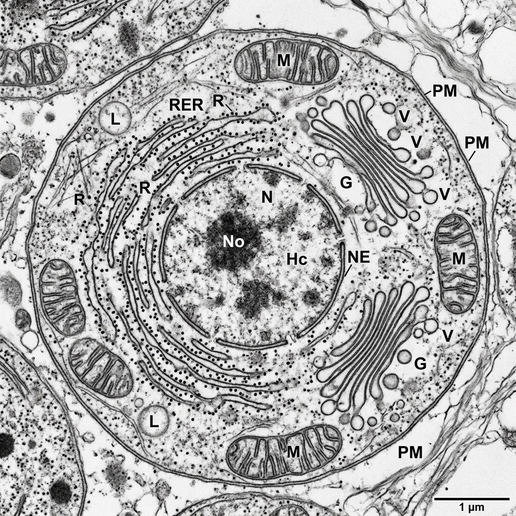
Skill: Drawing & Annotation
Practice identifying and drawing cell structures from electron micrographs (like the one above). Key structures to identify:
- Nucleus: Large dark circular region (often with a darker nucleolus).
- Mitochondria: Oval structures with inner folded membranes (cristae).
- Rough ER: Network of membranes studded with dark dots (ribosomes).
- Golgi Apparatus: Stack of flattened, curved sacs without ribosomes.
A2.2.12 HL Only
Endosymbiosis
Key Understanding (HL) The origin of eukaryotic cells is explained by the Endosymbiotic Theory.
The theory states that mitochondria and chloroplasts were once free-living prokaryotes that were engulfed by a larger host cell. Instead of being digested, they formed a mutually beneficial symbiotic relationship.
Large ancestral prokaryote engulfs smaller aerobic bacteria
↓
Symbiosis: Host gets ATP; bacteria gets protection
↓
Over millions of years, engulfed bacteria evolve into Mitochondria
Evidence (Mitochondria & Chloroplasts): They possess double membranes, their own naked circular DNA, 70S ribosomes, and replicate by binary fission independently of the cell!
A2.2.13 HL Only
Cell Differentiation
Key Understanding (HL) In multicellular organisms, cells differentiate to perform specialized functions by expressing some genes and not others.
Every somatic (body) cell in a multicellular organism contains the exact same genome (identical DNA). However, cells look and function differently (e.g., a neuron vs a muscle cell).
Gene Expression:
Differentiation occurs when specific genes are "turned on" (expressed) while others are "turned off" (repressed). Chemical gradients and environmental signals trigger this selective gene expression, directing the cell along a specific developmental pathway.
A2.2.14 HL Only
Evolution of Multicellularity
Key Understanding (HL) Multicellular organisms have emergent properties arising from the interaction of their cellular components.
As organisms grew larger and more complex, solitary cells grouped together. Over evolutionary time, this cooperation led to true multicellularity.
Cells group together (Colonies)
↓
Cells differentiate and specialize (Tissues)
↓
Tissues work together for complex functions (Organs)
↓
Emergent Properties: The whole organism can do things that the individual cells cannot do alone.
✅
A2.2 Cell Structure Complete!
You have successfully reviewed all SL and HL understandings for Biology Theme A2.2.