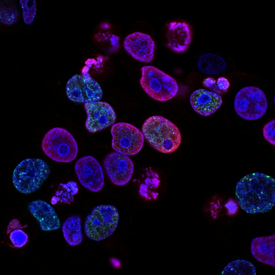
Blood cells and heart imagery

B3.2 — Transport

Theme B: Organismal Level · Standard & Higher Level

10
Understandings
8
HL Extensions
B3.2.1 - B3.2.3

Blood Vessels: Arteries, Veins, and Capillaries

Key Understanding Arteries convey blood at high pressure from the ventricles to the tissues. Veins return blood to the atria. Capillaries exchange materials.

The mammalian transport system relies on three distinct types of blood vessels, each structurally and functionally adapted to ensure the efficient transport of respiratory gases, nutrients, and waste products across the body.
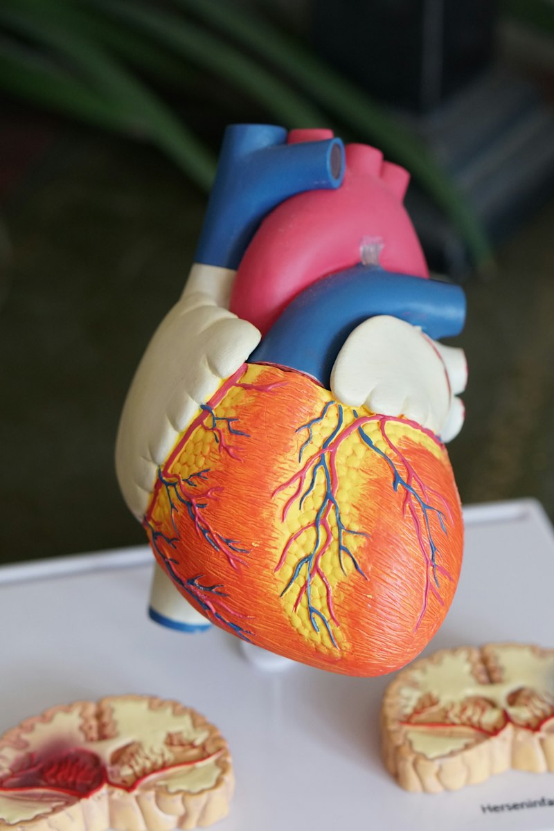
Microscopic view of blood vessels Red blood cells flowing

Arteries

  • Transport oxygenated blood (except pulmonary artery) away from the heart.
  • Possess thick, highly muscular, and elastic walls containing elastin fibers to withstand and maintain high hydrostatic pressure.
  • Narrow lumen ensures the high pressure of the blood is sustained between heartbeats.
  • Elastic recoil helps propel blood forward in pulses.

Veins

  • Transport deoxygenated blood (except pulmonary vein) towards the heart.
  • Have relatively thin walls with fewer muscle and elastic fibers, accommodating lower pressure blood flow.
  • Feature a much larger lumen to reduce resistance to flow.
  • Contain pocket valves to prevent the backflow of blood, especially against gravity (e.g., in the legs).
Capillaries

Capillaries are the microscopic site of material exchange (e.g., O₂, CO₂, glucose, urea). Their extraordinary adaptations include:

Vocabulary Check

Essential Terms: Human Transport

Hover over (or tap) each card to reveal the definition of these crucial cardiovascular terms.

Lumen (Hover to flip)
The central cavity or opening within a tubular structure, such as a blood vessel, through which blood flows.
Endothelium (Hover to flip)
The single layer of flattened cells lining the inner surface of all blood vessels, providing a smooth surface that reduces friction.
Systole (Hover to flip)
The phase of the heartbeat when the heart muscle contracts and pumps blood from the chambers into the arteries.
Diastole (Hover to flip)
The phase of the heartbeat when the heart muscle relaxes, allowing the chambers to fill with blood.
B3.2.4 & B3.2.6

Pulse Rates and Coronary Heart Disease

Skill: B3.2.4 Measurement of Pulse Rates

The pulse can be palpated in superficial arteries (e.g., the radial artery in the wrist or the carotid artery in the neck). It is a direct result of the elastic recoil of arterial walls immediately following the ejection of blood from the left ventricle. Pulse rate is quantitatively measured in beats per minute (bpm). Students should master measuring it manually over a defined time interval or by utilizing modern digital plethysmography sensors to analyze heart rate variability.

Application: B3.2.6 Occlusion of Coronary Arteries

The heart muscle (myocardium) requires its own dedicated blood supply via the coronary arteries. When these arteries become occluded (blocked), it leads to coronary heart disease (CHD).

B3.2.7 - B3.2.8

Transpiration and Xylem Adaptations

Key Understanding Water is continuously transported from roots to leaves during transpiration due to the combined forces of cohesion, tension, and adhesion.
Plant biology diagram context Forest canopy showing transpiration context

The Transpiration Stream: This process describes the uninterrupted flow of water through a plant, driven primarily by solar energy causing evaporation.

Solar heat causes water to evaporate from spongy mesophyll cell surfaces and diffuse out of stomata as water vapor.
This evaporation creates a strong pulling force (tension or negative pressure, $\Psi < 0$) that draws water out of leaf xylem vessels.
Cohesion: Hydrogen bonds tightly associate H₂O molecules, maintaining a continuous, unbroken column of water down to the roots.
Adhesion: H₂O molecules form hydrogen bonds with the hydrophilic cellulose and lignin in xylem walls, resisting gravity.
Xylem Vessel Adaptations (B3.2.8)

Xylem vessels are exquisitely adapted for efficient, unidirectional mass flow of water under immense negative pressure:

B3.2.9 - B3.2.10

Tissue Distribution in Dicotyledonous Plants

Key Understanding Students must confidently identify the characteristic distribution of tissues in transverse micrographs of dicot stems and roots.

The spatial arrangement of vascular tissue (xylem and phloem) is distinctly adapted to resist the specific mechanical stresses experienced by different plant organs.

Dicotyledonous Stem

  • Vascular bundles are systematically arranged in a ring situated near the outer periphery (epidermis).
  • Within each bundle, the thick-walled xylem is located on the inside, while the phloem is positioned on the outside. A layer of cambium typically separates them.
  • Function: This peripheral ring structure provides maximum resistance to bending forces caused by wind, acting much like a hollow structural cylinder.

Dicotyledonous Root

  • Vascular tissue is condensed into a tight central cylinder known as the stele.
  • The xylem typically forms a central, robust star or cross shape.
  • The phloem is strategically located in the spaces between the arms of the xylem star.
  • An endodermis surrounds the stele, controlling water entry.
  • Function: The central arrangement is optimized to withstand strong vertical pulling forces, anchoring the plant firmly against upward tension.
B3.2.11 - B3.2.14 HL Only

Tissue Fluid and Circulation Types

Tissue Fluid Formation and Reabsorption (B3.2.11 - B3.2.13)

Capillaries exchange materials not directly with cells, but via an intermediary called tissue (interstitial) fluid.

Single Circulation (e.g., Fish)

  • Blood completes one full circuit through a two-chambered heart per systemic trip.
  • Pathway: Heart $\rightarrow$ Gills (oxygenation) $\rightarrow$ Systemic Capillaries (body) $\rightarrow$ Heart.
  • Passing through the delicate gill capillaries drastically reduces blood pressure, meaning blood flows relatively slowly to the rest of the body.

Double Circulation (e.g., Mammals)

  • Blood passes through a four-chambered heart twice per circuit, completely separating oxygenated and deoxygenated blood.
  • Pulmonary Circuit: Right ventricle $\rightarrow$ Lungs (low pressure, prevents lung damage).
  • Systemic Circuit: Left ventricle $\rightarrow$ Body (high pressure, ensuring rapid and efficient O₂ delivery to metabolically highly active tissues).
B3.2.15 - B3.2.16 HL Only

The Cardiac Cycle and Heart Adaptations

Key Understanding (HL) Deeply understand the anatomical adaptations of the heart chambers and the pressure/volume changes during the cardiac cycle.
Skill: B3.2.16 Cardiac Cycle and ECG Interpretation

The cardiac cycle comprises carefully coordinated phases of contraction (systole) and relaxation (diastole). An Electrocardiogram (ECG) visualizes the electrical depolarization and repolarization triggering these phases: the P wave (atrial depolarization), the QRS complex (ventricular depolarization), and the T wave (ventricular repolarization).

1. Atrial Systole: Atria contract simultaneously. Pressure in the atria momentarily exceeds ventricular pressure, forcing the remaining blood through open atrioventricular (AV) valves into the relaxed ventricles. Semilunar valves are closed.
2. Ventricular Systole: Ventricles contract forcefully from the apex upwards. Ventricular pressure rapidly spikes, snapping the AV valves shut (the first heart sound, "lub", preventing backflow). When ventricular pressure exceeds aortic/pulmonary pressure, the semilunar valves are forced open, ejecting blood into the arteries.
3. General Diastole: Ventricles and atria relax. Ventricular pressure plummets below arterial pressure, causing semilunar valves to snap shut ("dup"). Once ventricular pressure drops below atrial pressure, AV valves passively open, and blood flowing from veins begins refilling the heart.
B3.2.17 - B3.2.18 HL Only

Root Pressure and Phloem Translocation

B3.2.17 Root Pressure

In certain conditions (high humidity, low transpiration), plants can still push water upwards. Root cells utilize ATP to actively transport mineral ions from the soil into the root xylem. This massive solute accumulation drastically lowers the water potential ($\Psi$) inside the xylem. Consequently, water rapidly enters from the surrounding root cortex via osmosis, generating a strong positive hydrostatic pressure (root pressure) that physically pushes the sap column up the stem, occasionally manifesting as guttation (water droplets on leaf margins).

Phloem Adaptations for Translocation (B3.2.18)

The phloem actively transports dissolved organic compounds (primarily sucrose and amino acids) from sources (e.g., mature leaves) to sinks (e.g., growing roots, fruits) via a mechanism explained by the pressure-flow hypothesis.

Self-Assessment

Check Your Understanding

Expand the questions below to verify your mastery of the B3.2 Transport module concepts.

1. Contrast the structural adaptations of arteries and veins and explain how they relate to their functions.

Arteries possess thick, highly muscular walls with abundant elastic fibers. This allows them to withstand and actively maintain the high hydrostatic pressure of blood freshly pumped from the heart. Their narrow lumen aids in maintaining this pressure. In contrast, veins have thinner walls with fewer muscle and elastic fibers, accommodating lower pressure blood returning to the heart. They feature a larger lumen to reduce resistance to flow and incorporate pocket valves along their length to prevent the backflow of blood against gravity.

2. Explain the mechanism by which water is transported against gravity from roots to leaves in a tall tree.

Water transport is driven by transpiration. Solar heat causes water to evaporate from the spongy mesophyll and exit via stomata. This creates a negative pressure (tension) in the leaf xylem. Because water molecules are highly polar, they exhibit cohesion (bonding to each other via hydrogen bonds) and adhesion (bonding to the hydrophilic lignin in xylem walls). The tension from the leaves pulls the continuous, unbroken column of water up the xylem vessels from the roots to replace the lost water.

3. Describe how tissue fluid is formed at the capillary bed and how it is reabsorbed. [HL Only]

At the arteriole end of a capillary, high hydrostatic pressure from the heart forces water and small solutes (like glucose and oxygen) out through the endothelial fenestrations (ultrafiltration), forming tissue fluid. Large proteins remain in the blood. As blood reaches the venule end, the hydrostatic pressure is much lower. However, the concentrated plasma proteins create a high osmotic pressure. This osmotic pull draws water and cellular waste products back into the capillary. Any excess fluid is drained by the lymphatic system.

4. Outline the events of ventricular systole in the mammalian cardiac cycle. [HL Only]

Ventricular systole begins when the electrical signal from the AV node spreads through the Purkinje fibers, causing the thick muscular walls of the ventricles to contract powerfully from the apex upwards. The pressure inside the ventricles rapidly increases, exceeding the pressure in the atria, which snaps the atrioventricular (AV) valves shut (preventing backflow and creating the "lub" sound). As pressure continues to rise above the pressure in the aorta and pulmonary artery, the semilunar valves are forced open, and blood is ejected forcefully into the systemic and pulmonary circulations.

B3.2 Transport Complete!

You have successfully reviewed all SL and HL understandings for Biology Theme B3.2.